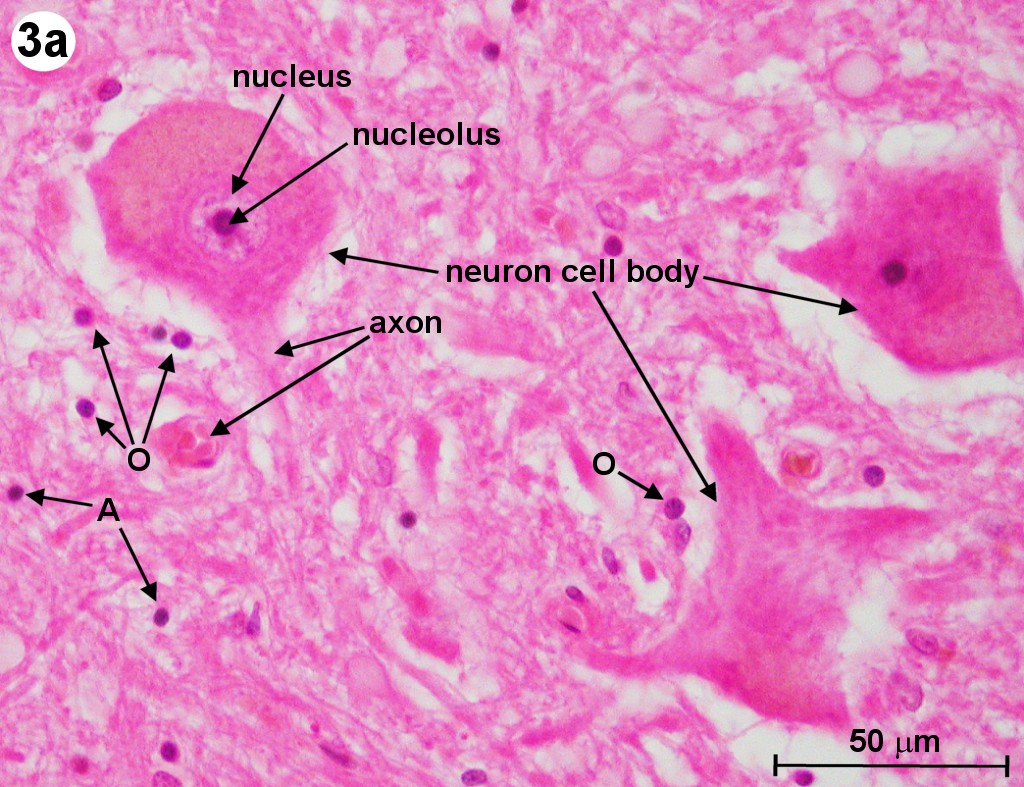
Label Each Region of the Neuron on the Image Below.
We are releasing this new dataset with our paper. Figure 1 C shows a photo-labeled neuron in this case a PLP PN white arrow among other neurons that also express PA-GFP but are not photo-labeled gray arrows.

Coronal View Ventricles Brain Diagram Brain Anatomy Corpus Callosum
Image segmentation is a computer vision task which involves labelling various regions of the image into objects that.

. Connecting different types of neurons D3. Nervous System - Neuron. It cannot be implemented real time as it takes around 47 seconds for each test image.
The human brain is estimated to contain over 100 billion neurons with each neuron having on average connections to 1000 other neurons. This neuron part sends on messages to other neurons. Identify the terms based upon the descriptions below.
The neuron labeled number _____ in the figure is a motor neuron. Connecting different types of neurons. Choose the correct names for the parts of the neuron.
Let Y i 2 f 11 g represent the binary labels indicating presence or absence of an obstacle at the ith location in the image and X i represent the input. Stimulating muscles or glands C1. Learn vocabulary terms and more with flashcards games and other study tools.
This is an image of a neuron. For each set of image regions we asked three distinct crowd workers to describe what they had in common resulting in nearly 60000 descriptions. This neuron part receives messages from other neurons.
If a secondary label is also imaged this image will be displayed alongside the primary image. What is the part labeled 1. The axon terminal is designed to convert the electrical signal into a chemical signal in a process called synaptic transmission further explained in the section Physiology of.
The pink lines that are attached to these neurons are their processes. We will produce a set of q predicted regions p k F such that the final predicted segmentation is P. RMP threshold depolarization repolarization hyperpolarization Propagation of Action Potentials 33.
Stimulating muscles or glands B3. _____ _____ occurs in a myelinated neuron. The list below is what happens when a neuron fires and sends a signal along to another neuron.
Label the following parts of the synapse and describe what it is what it does. This vast connectional architecture explains that computational complexity of the human brain. More formally an MRF is an undirected graph G V E where we represent each labeled location in the image as a vertex V and edges E connecting neigh-boring image locations.
We computed the top-activating image regions for 17000 neurons across 7 models which themselves span several architectures and tasks. Its function is _____. Next a selected person must manually label each image into several categories without having any information on the neurons activations.
Label A Neuron Worksheet - Weavingaweb The cell body of the neuron. BYJUS Online learning Programs For K3 K10 K12 NEET. Draw and label a neuron.
It takes a huge amount of time to train the network as you would have to classify 2000 region proposals per image. The entire morphology of this photo-labeled neuron is clearly visible including its dendrites axonal projection and presynaptic boutons Figure 1 C yellow. Axon is a tube-like structure that carries electrical impulse from the cell body to the axon terminals that passes the impulse to another neuron.
Label each region of the neuron on the image below. Sodium ions diffuse through leakage channels into the cell but potassium ions diffuse through leakage channels out of the cell. This neuron part processes incoming messages.
Once an axon reaches a target it terminates into multiple endings called axon terminals. The Diagram Below Is Of A Nerve Cell Or Neurone. This neuron part contains instructions for making.
Consider an image volume consisting of m image slices that is to be segmented into a set of n true regions t l such that the true segmentation is t 1 t 2 t nEach t l in has a unique integer label and consists of a set of pixels v ijk that are 26-connected. Neurons communicate with each other by generating and conducting electrical signals. Each neuron has a cell body with a nucleus Golgi body endoplasmic reticulum mitochondria and other components.
If you like colour in the diagram as suggested below. Fill in the missing blanks in each statement. About 90 of the cells in the central nervous system--brain and spinal cord--are supporting cells sc.
Label color the different parts of the neuron below. As shown below because activations have an arbitrary scale the resulting conditional probability plots the activation axis in terms of standard deviations of activation from zero. The aim of semantic image segmentation is to classify each pixel of an image.
Image shows Anterograde and Retrograde transport in an axon. If the neuron contains regions of self-fasciculation the Length. This neuron part gives messages to muscle tissue.
Consider this main learning worksheet to teach all about the fabulous neuron cells and the 6 parts. On the image below label the correct location on the graph where the following events are occurring. Motor Neuron smear Each neuron n has extensions called processes axons and dendrites that allow it to communicate with other neurons.
Presynaptic Neuron Vesicles Calcium Channels Synaptic Cleft Receptor Molecule and Postsynaptic Neuron. Start studying Label Parts of a Neuron. Normally sodium and potassium leakage channels differ because ___________________.
The Neuron ROI NoiseROIs optional and Click Cell Body modules run sequentially on each displayed neuron image allowing the user to circle the neuron of interest circle. Science Biology QA Library Consider the image.

The Nervous System Is The Sensory And Control Apparatus Consisting Of A Network Of Nerve Cells Peripheral Nervous System Nervous System Diagram Sciatic Nerve

4 Main Parts Of The Brain And Their Functions Explained Brain Lobes Human Brain Brain Diagram
Biochemistry 9th Edition By Mary K Campbell Shawn O Farrell Owen M Mcdougalisbn 13 978 1305961135isbn 10 1305961137i Biochemistry Mary K Online Textbook
How Aphasia Causes Difficulty Speaking Brain Diagram Brain Anatomy And Function Aphasia

Image Result For Primary Somatosensory Cortex Cerebrum Brain Anatomy Somatosensory Cortex Brain Structure

Caudate Nucleus Google Leit Caudate Nucleus Basal Ganglia Basal Ganglia Stroke

The Spinal Cord Organization Of The Central Nervous System Part 1 Spinal Cord Nervous System Parts Spinal

Ap Bio Nervous System And Muscles Brain Anatomy And Function Brain Nervous System Occipital Lobe

Aphasia Amy Speech Language Therapy Inc Aphasia Brain Models Aphasia Therapy
No comments for "Label Each Region of the Neuron on the Image Below."
Post a Comment